
Neurotransmitter overview
As Andrawis (2018) has pointed out, neurotransmitters are brain messengers consisting of biochemical substances that serve to transfer stimuli from one cell to another through the synaptic connection. They are produced in the cell body or in the axon of the conducting neuron and released to a certain extent. The human brain has more than 100 billion nerve cells that communicate with each other.
Cooperation takes place, on the one hand, through the transmission of information through electrical impulses that are transmitted and, on the other hand, through biochemical processes that are collectively referred to as neurotransmitters. Neurotransmitters have the task of conducting information between the nerve cells, with the synapses playing a role as contact points. This transmission takes a few milliseconds. The neurotransmitters consist of five different interacting messengers, the important neurotransmitters that play a role in mental illness:
I. Acetylcholine Glutamate and Gamma-Aminobutyric Acid (GABA)
II. Dopamine
III. Serotonin
IV. Adrenaline
V. Noradrenaline.
These are specific substances in which neurons are present and synthesized (Andrawis A, 2018). The presynaptic neurons are released there in their end region. Classic neurotransmitters are dopamine, serotonin, acetylcholine, norepinephrine, as well as gamma-aminobutyric acid, aspartate, glutamate, homocysteine, glycine and taurine. There are also other transmitters that may not meet these criteria, e.g. ACTH, endorphins, peptides, cholecystokinin. These are synthesized in the cell body and not in synaptic molecules and later they are transported to the synapses. The neurotransmitter peptides have the same function as the classical neurotransmitters. Together they have a regulating and modelling function. Several hundred peptides and neurotransmitters have been found in neuroscience (ibid.).
The pre- and post-synaptic neurotransmitters are the place where the excitation transmission of the neurotransmitters and their biochemical processes take place. Due to the effect of acetylcholine on the postganglionic sympathetic neurons, a “depolarized state” is created by electrical discharge, which triggers the action potential. This in turn triggers an interaction between the action potentials. The speed of an electrical nerve impulse is 1m/s and follows along the axion to the nerve end, continues to the sympathetic innervated to the target organs. This leads to a release of noradrenaline, which binds transmission substances from the neural storage sites to the noradrenergic and adrenergic receptors in the target organs. Vascular constriction is triggered by noradrenergic receptors, e.g. when noradrenergic vascular muscles are reached (ibid.).
The lifestyle and the experiences and events associated with it lead to an activation of genes and thus to a change in the brain structure. Situational influences have an effect on the regulation of gene activities. The gene regulates circulation, hormones, blood sugar and stress regulation, while the immune infection and tumor defense also depend on the regulation of genes. The metabolic processes are regulated by proteins and controlled by the gene. It reads a section of a sequence and its contents from the DNA genetic material and processes this information in small steps so that protein can be built up and converted.
The human genes, which have over 35,000 DNA sequences, are largely identical from person to person, but they exhibit individual genetic patterns of ethnic and constitutional differences. The gene text only appears altered in 0.1% of the population. These are hereditary diseases such as cystic fibrosis or cholera. However, genetic diseases are very rare, only 6% of the population have Alzheimer’s and 2% genetic mutations (ibid.).
Acetylcholine molecules
Acetylcholine primarily mediates nerve impulses to the periphery of the musculature. The messenger substance also plays an important role in controlling the autonomic nervous system, e.g. heartbeat, respiration and metabolic control.
In less than one thousandth of a second, acetylcholine molecules enter the cell membrane via the synaptic cleft. There, a specific cell membrane post-synaptic molecule is bound from protein molecules to sympathetic neurons, resulting in a specific molecule of acetylcholine receptors that fit together. When the receptors are occupied and polarized with the specific protein, the postganglionic neurons are excited and activated. The electrical charge of the membrane releases negatively charged electrical molecules on the outside of the membrane and positively charged molecules on the inside. The sleep state is now reached. Furthermore, Klußmann & Nickel (2009) describe that certain gene activities depend on the synaptic excitation of nerve cell transport. In extraordinary life situations these react simultaneously with the synaptic activity. By alerting the nerve-damaging messenger substance glutamate, gene production is increasingly stimulated (ibid.).

Fig.1 Acetylcholine molecules
Source: https://bit.ly/2IPUt9Z
Acetylcholin belongs to the chemical substances of a neurotransmitter. Its production takes place in the nucleus accumbens, the striatum and the olfactory tubercules. It also reaches cholinergic endings up to the substantia innominata. The transferase of acetylcholine with acetylkoenzymes and acholine causes that choline cannot be synthesized. It enters the bloodstream via food and is stored, synthesized and absorbed in the presynaptic nerve endings after its formation. The cholinergic system plays the most important functional role between the nerve and muscle cells of the end plate. In the CNS, a binding with dopaminergic systems takes place, which has been identified as a cure for Parkinson’s syndrome (Schmitz 1999).
The inhibitory transmitter GABA (y-aminobutyric acid) is found in the brain and spinal cord. This plays an important role in addictive behavior. Benzodiazepines, ethanol and barbiturates show a defective reaction at the components, which causes post-synaptic inhibition of the GABA system and at the macromolecular receptors and therefore puts the muscle into a sedating state (Andrawis A, 2018).
GABA neurotransmitters will carry 30% of the neurons. Two long, projecting neurons can be distinguished, which have extensive axons, as can be seen in the example of the Purkinje cells of the cerebellar cortex or the GABA-ergic neurons. The latter are present in a predominant amount of neurons, this is due to short interneurons. They play an essential role in excessive neuronal activities (ibid.).
Dopamine
Dopamine has a special significance for the control of the musculoskeletal system, which is manifested by arbitrary actions. It is located in the central nervous system and consists of two dopaminergic substances: The substantia-nigra and striatum. An excess of dopamine leads to mental illness, stiffness and trembling of the muscles. Dopamine deficiency reduces movement impulses and can also lead to Parkinson’s disease. It is still unclear whether dopaminergic overactivity is the cause of this disorder. However, it is assumed that dopamine excess or hypersensitivity of dopaminergic receptors is the starting point. Möller suggests that this is an excess of dopaminergic activity in relation to other neurotransmitter systems (ibid.).
The serotonergic and glutamatergic systems play an important role in schizophrenia. This hypothesis is still being discussed today. Neuroleptics are the antagonists (dopamine D2 antagonists), they trigger acute symptoms and cause hallucinations. Amphetamines can also be the cause of increased dopamine transmission. The glutamaterge and the dopaminerge system are closely linked. The serotonergic system is also receiving increased attention, as all neuroleptics have a serotonin-5HT2A antagonism in addition to the aforementioned dopamine D2 antagonism. The psychosocial factors play an important role as cause and trigger of schizophrenia. This disease is more common in lower social classes. Stress caused by overstimulation also has a negative effect on the development of this disease (ibid.).
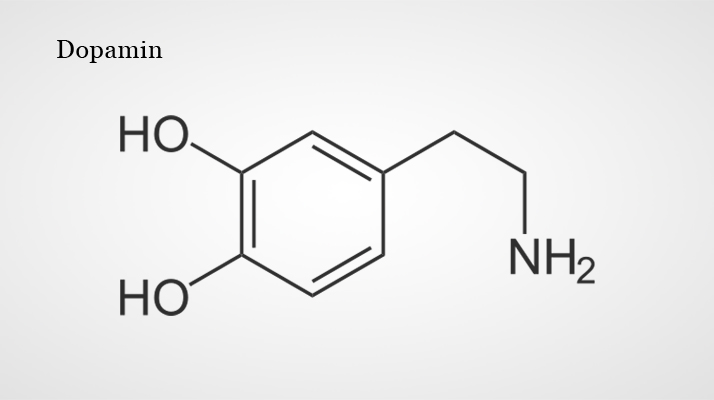
Fig. 2 Dopanim
Source: https://bit.ly/2HhsnaJ https://bit.ly/2HhsnaJ (19.04.2018 16:04)
Fig. 2: shows the dopaminergic railway systems with the different target group systems in the CNS.
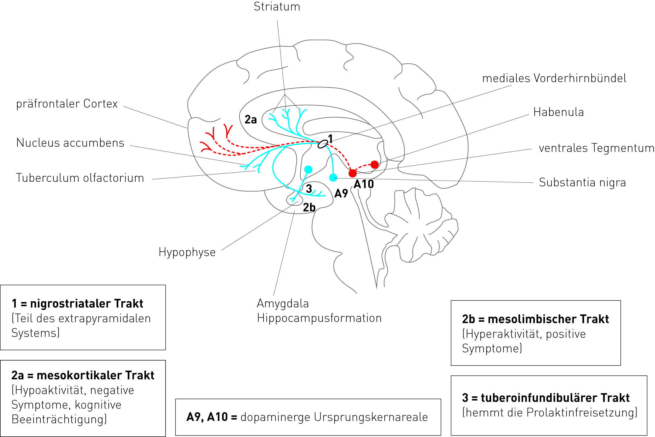
Fig. 3: Dopaminergic systems
Source: (Möller et al. 2005:138)
The tasks within dopaminergic systems are divided into different cell group systems: mesolimbic, mesostriatic, mesocortical and hypothalamic. The neurons of the mesolimbic and mesocortical systems are projected into the cortical region, the limbic system and in the septum to the pyriform, amygdalakerne and locus coeruleus (Andrawis A, 2018).
The mesostriatal system consists of a dorsal and a ventral part. The dorsal part provides dopamine to the mesostriatal system, carries neurons to the striatal regions, as well as putamine, gaudatus and globus pallidus further into the subthalamic neurocortex and nucleus. The ventral part belongs to the retrorubral core, which is produced. The mesostrial, dopaminergic system plays a major role in motor movements: In the event of a disorder occurring, motor disorders and Parkinson diseases result. Schmitz explains that locus coeruleus and neurons of the lateral segmentum are the origin of most noradrenergic systems. Receptors include G proteins and second messenger systems. A distinction is made between α and β receptors. Two subtypes also contain a1 receptors for C receptors for the release of infracellular calcium and phospholipase (ibid.).
Serotonin
Serotonin influences the sleep-wake rhythm, mental state, pain perception and blood pressure. At higher concentrations, vigilance is active, but can also cause restlessness and hallucinations. The lack of serotonin triggers aggression, anxiety and depression. Biogenic amines include serotonin, dopamine and norepinephrine. The amino acid tryptophan is synthesized from them. The enzyme tryptophan hydroxylase oxidizes to 5-hydroxytryptophan. The 5-hydroxytryptophandecarboxylase converts it into the substance 5-hydroxytryptamine(=5-HAT=serotonin). Serotonin and catecholamines are stored in the granules of the nerve endings and released by a calcium-dependent mechanism. These are actively resumed in the presynaptic cell and reach the synaptic cleft again after release. Serotonin is degraded via monoaminooxidase (MAO). This oxidation produces the product 5-hydroxyndolacetic acid (5-HIAA) (ibid. ).
Adrenaline
Adrenaline and noradrenaline are formed in the adrenal medulla and are both considered stress hormones. Adrenaline regulates muscle energies in the form of glucose and plays an important role in dangerous situations as well as in sporting performance (Rüegg 2003). The most important neurotransmitters are divided into small molecules and neuroactive peptides. Biogenic amines are adrenaline, dopamine, noradrenaline and histamine. Neuroactive peptides include opioids, tachykinins, insulins, neurohypophysials.
Most of the noradrenergic neurons are responsible for and starting point for two nucleins in the brain stem, consisting of Locus coerolus and neurons of the lateral segmentum. The Locus coerolus shows little neuronal activity when eating, sleeping, etc. As soon as new stimuli are offered, his activity increases. Massive anxiety and loss of pleasure sensations in atypical depressive patients may be attributed to a disorder of vegetative function in anxiety disorders. Thus, the effect of monoamine oxidase inhibitors (MAO inhibitors) and antidepressants, which can suppress the activity in animal experiments, can also be explained.
Like dopamine, noradrenaline is a catecholamine, which is synthesized via the L-dopa and dopamine stages. Catechol-O-methyltransferase (COMT) can also inactivate noradrenalin.
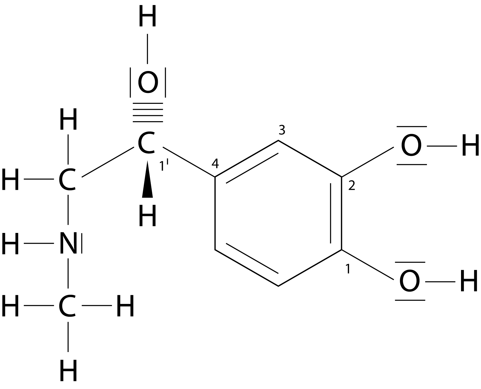
Fig. 4: Adrenaline
Source: (Löffler 2001: 481)
Noradrenergic System
Das noradrenerge System steuert die Aufmerksamkeit und deren Effekte und dient als phasisches System, das eine wichtige Rolle bei Störungen der vegetativen Funktion spielt. Es besitzt eine allgemein modulierende Funktion durch Lenkung der Aufmerksamkeit und Orientierung in Richtung neuer Stimuli, wo es eine ausführende Rolle hat, indem es parallel im peripheren sympathischen System sowohl die Aktivität, als auch die kortikale Informationsvereinbarung durch Signalrauschabstand diverser Systeme verändert (Andrawis A, 2018).

Fig. 5: Noradrenaline
Source: https://bit.ly/2HClrmp
The Noradrenalin
Noradrenaline is one of the most important neurotransmitters in the brain and is regarded as a stress hormone in the catecholamine group. The amino acid L-tyrosine is a component. The effect of noradrenalin is formed in the locus caeruleus. Released into the organism, it causes flight reflexes. The release of noradrenalin is induced by the adrenergic neurons which are CNS, thalamus, spinal cord, locus coeruleus and cerebellum in the central nervous system. The most important tasks include increasing attention and regulating behaviour in situations of fear and danger (ibid.). Norepinephrine is a neurotransmitter, a carrier substance like norepinephrine, which accelerates the heartbeat, for example when you get upset. In this way we can express our feelings or explain how the brain and psyche affect the heart and circulation.
Fainting or psychosocial stress are two examples. It is also described as an inner state of hopelessness, for example not being able to meet the demands of private and professional life. Mental stress already causes an increased circulatory disorder of the heart muscle, especially in sick people. Arbitrary and involuntary tension of the arm muscles is also not unproblematic, because the strong activation of the sympathetic nervous system drastically increases the frequency of the electrical impulses and action potentials, which reach the smooth muscles of the blood vessels, after they reach the heart in volleys fired from the nerve cells of the sympathetic nervous system. Action potentials triggered in the cell body are depolarized or electrically “discharged” by postgangliular sympathetic neurons through the action of acetylcholine, transferred from the cell body soma to the target organs and there cause the release of the carrier substance norepinephrine in the nerve storage sites, finally forming the adrenergic or noradrenergic receptors of the target organs and thus trigger a noradrenergic organ reaction such as vascular constriction, which is ultimately also the reason why noradrenalin enters the bloodstream and reaches the vascular muscles (ibid.).
Brain and serotonin systems
Excessive stimuli are modulated on a large scale. This explains the seemingly essential role of serotonin in homeostasis. The serotonin system can be explained in its protective function against feelings such as fear, helplessness and depression, which is shown by an inhibitory influence of negative feelings in behaviour.
Fig. 6: Serotonin
Source: https://bit.ly/2GWYuNC (22.04.2018 21:59)https://bit.ly/2GWYuNC (22.04.2018 21:59)
Directly or via stimulation, serotonin usually has an inhibitory effect on the GABA-genic, inhibitory system. 5-HT plays a central role in the mania or etiology of depression: It seems to play a direct or indirect role in the regulation of autonomous processes such as sleep, sexual activity, appetite regulation, circadian rhythms, body temperature, anxiety and motor and cognitive functions. For example, a tryptophan-free diet was suggested to a normal volunteer. This revealed slight depressive moods compared to tryptophan overabundance (Schmitz 1999). In the 1950s, the Rauwolfia alkaloid reserpine was prescribed for the treatment of high blood pressure. In repeated treatment, 15% of patients showed depressive symptoms. Later discoveries showed that dopamine, norepinephrine and serotonin led to an impoverishment of biogenic amines in the brain during reserpine treatment. The result was a release of the transmitters from the vesicles into the cytoplasm. They could then be distributed in the synaptic cleft. In 1965, Schildkraut formulated the catecholamine hypothesis regarding depression: The increase of the transmitter concentration causes an inhibition of the degrading enzyme MAO. This therefore antagonizes the effect of reserpine in animal experiments. Studies also showed that tricyclic antidepressants and Mao inhibitors played a major role in the treatment of patients with both noradrenaline and serotonin (ibid.).
In the case of drug abuse, the mesocorticolimbic dopamine system is more pronounced as a result of the amplification and reward system. This intensifies emotional functions of the brain and also leads to an increased motivation to act. Its functions are not only limited to activation and motivation, but also to the functional coupling of attention. This can be seen, for example, in learning and memory processes, in which learning process, conditioning and general memory performance were improved (ibid.). The following figure shows the structure of a nerve cell and its axon.
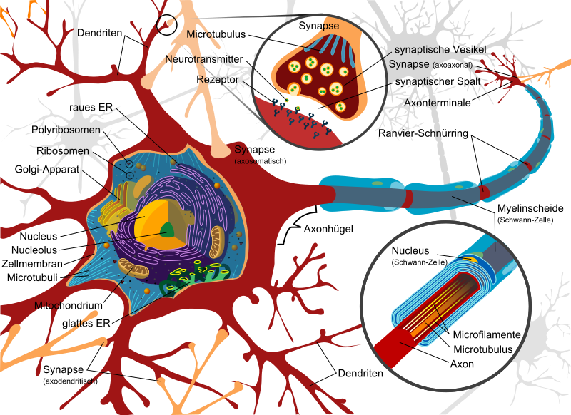
Fig. 7: Structure of a nerve cell and its axon
Source: https://bit.Ly/2BnHvg5 (22.04.2018 22:00)
Pre- and postganglionic neurons
The pre- and postganglionic neurons are connected to the sympathetic ganglia. The synapses consist of two cell membranes, with the binding neurons and the adjacent microscopically small synaptic cleft. The membrane in front of the gap is called presynaptic ganglion, the other membrane postsynaptic ganglion. The transmission of acetylcholine is released by the excitation of electrical impulses. This process is referred to as action potential (ibid.).
The Neuron
The neuron is usually associated with cell bodies, dendrites and an axon, as well as presynaptic area endings and has different functions, such as the transmission of signals.
In order to combine certain tasks, the electrical single-phase course is necessary to determine exact steps as well as the conduction of the bypass signal. This signal transmitter leads to specific regions of the neuron. These are dependent on electrical bases in the cell membrane (ibid.).
As Schmitz has described, exact neuronal communication takes place through connections between synapses and neurons. Although a neuron has about 1000 synaptic connections on average, this number can also vary, which is why we assume the input number 1011. If this starting point is reached, the synaptic number is increased to 1014 and formed in the brain. The information between the neurons is transmitted in two different ways: an electrical and a chemical compound (ibid.). Furthermore, according to Schmitz, the chemical neurotransmission of all neurons takes place in the same steps, while neurotransmitters are synthesized via presynaptic capsules. The storage is taken over by vesicles and released on a signal in the synaptic cleft. The cells communicate with each other through low-molecular compounds. The destination of these signal transducers is the receptors (ibid.).
Univ. Prof. Dr. Andrawis

Klicke hier, um Ihren eigenen Text einzufügen